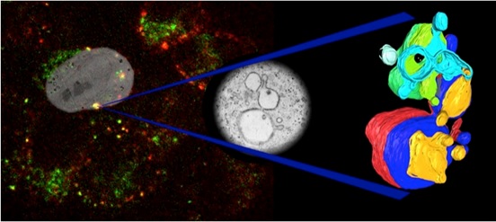
Correlative light electron microscopy (CLEM)
The Wolfson Bioimaging Facility is specifically equipped to combine light and electron microscopy and to undertake CLEM experiments. By utilising the advantages of fluorescence microscopy (e.g. live cell imaging) with the higher resolution and contextual information which EM provides, CLEM can deliver considerably more information than either modality alone.
Recent papers including CLEM data:
- Lee, Mantell, Hodgson, Alibhai, Fletcher, Brown, Frank, Xue, Verkade, Woolfson, Warren (2018) Engineered synthetic scaffolds for organizing proteins within the bacterial cytoplasm. Nat. Chem. Biol. 14:142-147
- Galloway, Senior, Fletcher, Beesley, Hodgson, Harniman, Mantell, Coombs, Rhys, Xue, Mosayebi, Linden, Liverpool, Curnow, Verkade, Woolfson (2018) Bioinspired silicification reveals structural detail in self-sssembled peptide cages. ACS Nano. 12: 1420-1432.
- Lees, Peddie, Collinson, Ashby, Verkade (2017) Correlative two-photon and serial block face scanning electron microscopy in neuronal tissue using 3D near-infrared branding maps. Methods Cell Biol. 140: 245-276.