Sample preparation laboratories
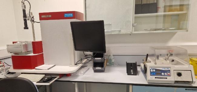
Polishing laboratory
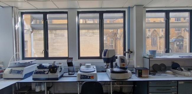
The Polishing Lab is located in Inner Court Laboratories and is available to anyone within the School of Earth Sciences. An induction and equipment/process training is required before access will be given (external use may be possible by arrangement).
The lab is well set up for polished resin block and thin section preparation, we have the following equipment;
- Buehler Ecomet /AutoMet auto-polisher
- Buehler MetaServ auto-grinder/polisher
- Buehler VibroMet vibratory polisher for ultra-fine polishing
- Buehler PetroThin thin-sectioning machine
- Buehler Isomet 1000 low-speed saw
- WELL Diamond Wire Saw
- Manual polishing and grinding equipment
- Sample bonding jigs and hot plates
- Reflected and transmitted light microscopes, with polarisers
- Stereomicroscopes for sample mounting and picking
- Heavy liquid separation and sample filtration apparatus
- Vacuum impregnation apparatus
- Technoorg Linda SEMPrep2™ ion polishing mill, which uses a beam of Ar+ions to polish samples to optimise electron microbeam analysis.
Rock crushing laboratory
The rock crushing lab is located in the basement of Wills Memorial Building, access is only granted after compulsory safety training and the completion of either a face-fit test for an FFP3 dust mask or training in the use of the positive pressure mask system.
The lab is equipped with a specialist dust extraction system and contains the following equipment;
- Retsch jaw-crusher
- Retsch PM100 planetary ball mill
- 15 tonne Hydraulic rock splitter
- 2 Dry vibratory sieve shakers
- 1 wet vibratory sieve shaker system
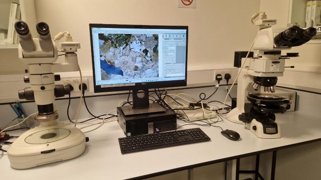
Optical microscopy laboratory
The optical microscopy lab houses a variety of microscope systems for the acquisition of high quality reflected and transmitted light images at high magnification. We have a dedicated PC with imaging software (NIS Elements) which also features image stitching.
- Nikon LV100N plain and polarised, transmitted and reflected light microscope
- Optional mechanised point-counting stage
- Nikon SMZ1000 stereomicroscope, with optional polariser
- SMZ1270i stereo microscope
All three microscopes have attached CCD cameras for the acquisition of petrographic images of thin sections and other geological samples.