Retinal neural progenitor cell
The neural progenitor cell group reside within the Academic Unit of Ophthalmology as part of an increasing sized and multi-collaborative research group which is working on progenitor cells within the retina. Within the last 5 years we have identified progenitor cells within the adult human retina. Our research is contributing nationally to a major translational research project on adult projenitor cells and represents the only group that has established this within adult post mortem neural retina.We are unique in that we are able to work with both donated human tissue and comparative work with animals (based in the Dorothy Hodgkin building).
Our research has to date established that within the adult human retina there exists a small population of cells that have a phenotype and characteristics of progenitor cells. We have purified these cells from retinal tissue using the stem cell marker CD133+ and have shown that they are capable of neurosphere formation, cell proliferation and cell differentiation into cell types found within the retina including glia, photoreceptors and neurons. Our aim is to achieve a greater understanding of the control of progenitor cells within the adult human retina within the normal and diseased retinal microenvironment. This will help form the basis of therapeutic treatment toward retinal regeneration. This may be in the form of (i) cellular therapies by replacing neurons/photoreceptor via transplantation of lineage specific stem cells or more significantly (ii) modulating micro environment to promote cell replacement, neuroprotection and prevent degeneration of neurons/photoreceptors.
Neurospheres express retinal cell and stem cell markers
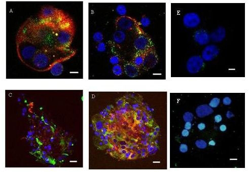 Confocal Images of Neurospheres Generated from CD133+ retinal cells cultured in FGF2/N2 A Neurosphere expressing Nestin+ (red) and Doublecortin+ cells (green X630) B Neurosphere expressing neurofilament+ (red) and doublecortin+ cells (green) X630. C Neurosphere expressing Nestin+ (red) and GFAP+ cells (green X400). D Neurosphere expressing Nestin+ (red) and rhodopsin+ cells (green X400). Nuclei counterstained with DAPI (blue). Neurospheres stained with isotype control antibodies did not have any significant fluorescent staining (E, X630, F, X630). Scale bars represent 5µm (A, B, E) and 10 µm (C, D, F).