|
This is not even a cursory look at nervous tissue, but it does cover most of the sections that you will encounter in the practical class.
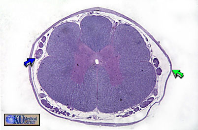 |
Figure 15 - Spinal Cord in transverse section. |
In the spinal cord, the gray matter is found in the center (the butterfly shape) and the white matter surrounds it. The opposite arrangement is found in the cortex of the brain. Notice the dorsal root (blue arrow) and the surrounding dura mater (green arrow).
===========================================================
 |
Figure 16 - White Matter. Bar is 50 microns |
The white matter of the spinal cord consists of myelinated fibers. Notice the numerous fibres surrounded by clear space where the myelin was prior to tissue processing.
===========================================================
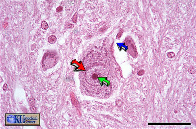 |
Figure 17 - Gray Matter. Bar is 30 microns |
Gray matter within the spinal cord contains many nuclei such as this multipolar motor neuron. Notice the nucleolus (green arrow) within the nucleus (red arrow) and what appears to be fibre (blue arrow).
===========================================================
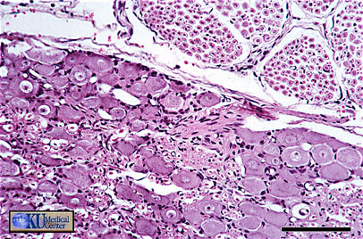 |
Figure 18 - Dorsal Root Ganglion (DRG). Bar is 100 microns |
Compare the large soma (perikarya) of the ganglion cells with the smaller myelinated nerves cut in cross section in the upper right. The DRG houses the cell bodies (somata) of the peripheral sensory nerves, although there are no synapses in the DRG.
===========================================================
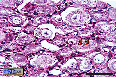 |
Figure 19 - Dorsal Root Ganglion. Bar is 50 microns |
At higher magnification view shows the surrounding satellite cells (red arrows), the glia of the DRG.
===========================================================
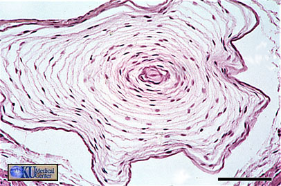 |
Figure 20 - Pacinian Corpuscle. Bar is 100 microns |
The Pacinian corpuscles are large "onion-like" encapsulated pressure receptors which you may happen to find when examining sections of GI tract. The surrounding concentric lamellae respond to distortion and generate an action potential in the bare nerve ending in the core.
|

