|
-
Central Nuclei
-
Cytoplasm to nuclear area ratio low
-
Relatively few cells sectioned through nucleus
 |
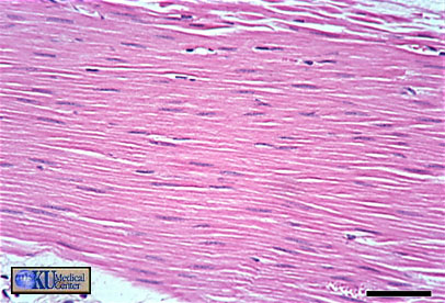
Figure 12 - Smooth muscle in longitudinal section. Bar is 50 microns |
Smooth muscle in longitudinal section is identified by long, thin central nuclei, a more scattered tissue layout, and an absence of striations.
===========================================================
 |
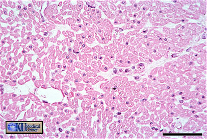
Figure 13 - Smooth muscle in transverse section. Bar is 250 microns |
Smooth muscle in transverse section can be identified by central nuclei and a small ring of surrounding cytoplasm. It may be evident that relatively few cells have been sectioned through the nucleus. Compare this to cardiac muscle in TS.
===========================================================
 |
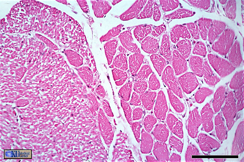
Figure 14 - Skeletal & smooth muscle in transverse section. Bar is 100 microns |
A closer look clearly shows skeletal muscle to the right and smooth muscle to the left.
|

