|
-
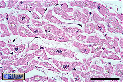
Central Nuclei
-
Cytoplasm to Nucleus area ratio is high
-
Nuclei are visible in many ventricular cells in transverse section (compared to very few nuclei in TS of smooth muscle)
 |
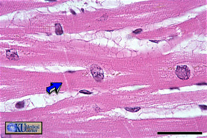
Figure 6 - Cardiac muscle in longitudinal section (bright field illumination). Bar is 30 microns |
Cardiac muscle in longitudinal section can be identified by centrally placed round to oblong nuclei, striations, branching, and intercalated discs (arrow)
===========================================================
 |
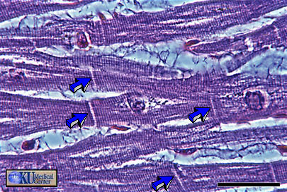
Figure 7 - Cardiac Muscle in longitudinal section (phase-contrast optics). Bar is 30 microns |
Compare this photomicrograph of the same slide as above (6), this time taken using phase-contrast optics. Notice how prominent the striations and intercalated disks (arrows) are when viewed using phase-contrast optics.
===========================================================
 |
Figure 8 - Cardiac muscle in transverse section. Bar is 50 microns |
In transverse section, cardiac muscle is identified by central nuclei and a large ring of surrounding cytoplasm. You may notice a high density of blood vessels and a relatively high proportion of cells that have been sectioned through the nucleus. These features would enable you to distinguish between cardiac and smooth muscle.
|

