Vectors, Clone
information and Methods
Adenoviral
vectors
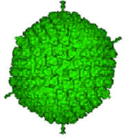 are produced using standard cloning techniques, followed by homologous
recombination according to a procedure adapted from Graham and Prevec (1994). High
purity viral stocks are then obtained using CsCl ultracentrifugation. This
yields replication-deficient and non-toxic vectors which are capable of
transgene transfer into neurones both in vivo and in vitro. Adenoviral
vectors are either microinjected into the relevant brain nuclei or used to
transfect organotypic slice cultures. Transgene expression is detectable from around
6h after infection, fully developed 48-72h and still taking place up to 3-4
weeks post infection.
are produced using standard cloning techniques, followed by homologous
recombination according to a procedure adapted from Graham and Prevec (1994). High
purity viral stocks are then obtained using CsCl ultracentrifugation. This
yields replication-deficient and non-toxic vectors which are capable of
transgene transfer into neurones both in vivo and in vitro. Adenoviral
vectors are either microinjected into the relevant brain nuclei or used to
transfect organotypic slice cultures. Transgene expression is detectable from around
6h after infection, fully developed 48-72h and still taking place up to 3-4
weeks post infection.
Lentiviral
vectors 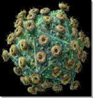 have been
recently established in this lab via a collaboration with Prof. M. Raizada'
group (University of Florida, USA). These vectors are based on the modified HIV
genome. They integrate into the host chromosome and therefore are able
to cause very long-term expression. For details see:
have been
recently established in this lab via a collaboration with Prof. M. Raizada'
group (University of Florida, USA). These vectors are based on the modified HIV
genome. They integrate into the host chromosome and therefore are able
to cause very long-term expression. For details see:
Efficient Large-Scale
Production and Concentration of HIV-1-Based Lentiviral Vectors For Use In
vivo
Coleman,J.E.;
Huentelman,M.J.; Kasparov,S.; Metcalfe,B.L.; Paton,J.F.R.; Katovich,M.J.;
Semple-Rowland,S.L.; Raizada,M.K.
Physiological
Genomics (2003), 12, 221-228
CLONES
LENTIVIRAL PLASMIDS (CLICK
TO DOWNLOAD, or Right-click and "save to folder". Open with Vector NTI or any
other software which reads *.gb files.
These files can be also open as text files to extract the sequences).
pTYF-2xGfaABC1D-tTA - "SuperGFAP
driving expression of Tet-off"
pTYF-2xSYN-tTA
- "SyperSYN1 driving expression of Tet-off"
pTYF-2xGfaABC1D-tTA - "SuperGFAP
driving expression of Tet-off"
pTYF-mCMS-SYN-EGFP -
(clone 238) "Bi-ridectional SYN1 driving expression of EGFP"
pTYF-mCMV-GfaABC1D-EGFP
- (clone 248)
"Bi-directional short GFAP driving expression of EGFP"
Information
about plasmids for
Lentivirus production
Organotypic
Brain Slices Cultures
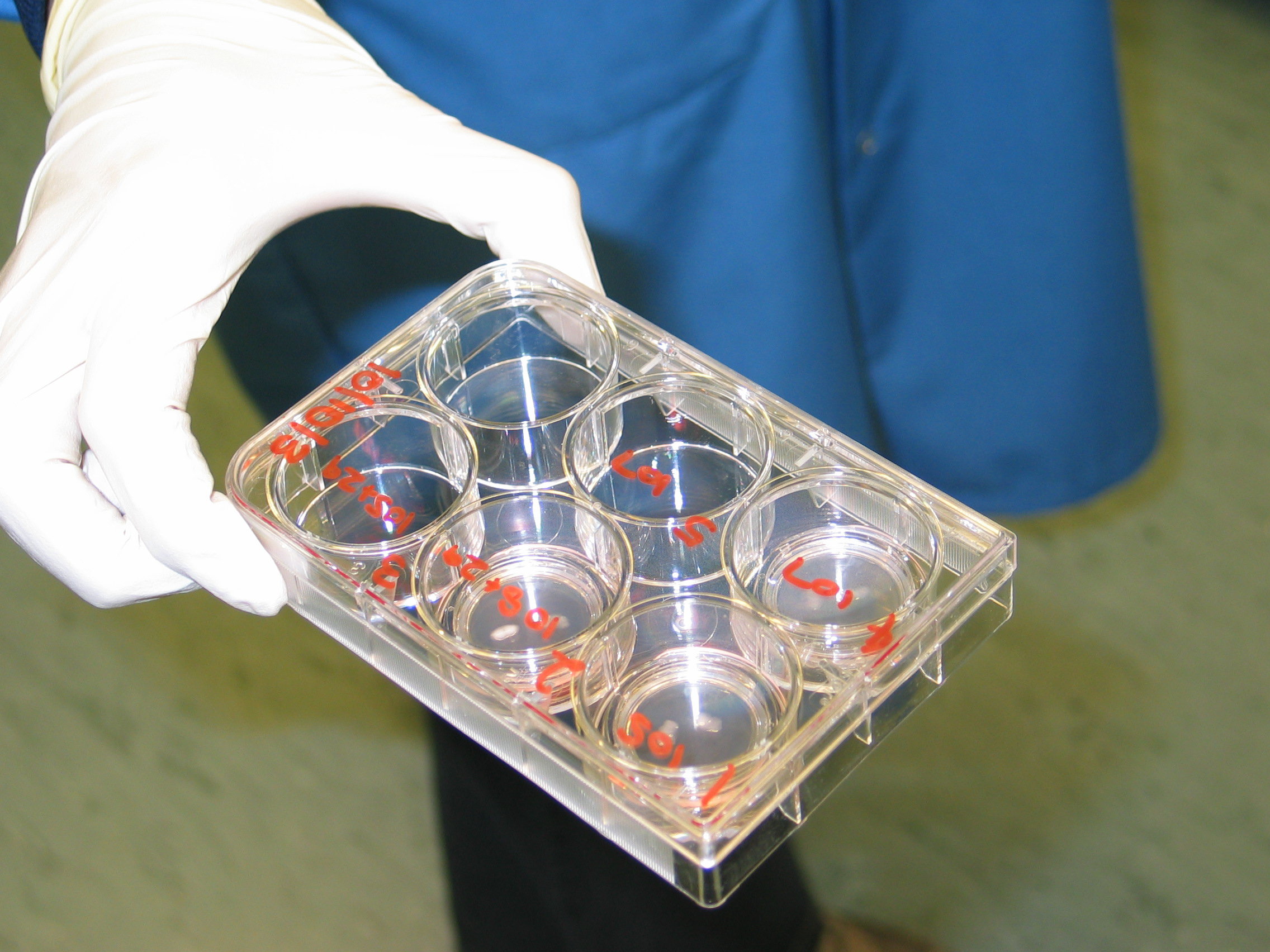
of
the rat brain(stem) are used as the main in vitro model to be combined with
viral transgenesis. They are prepared by the static culturing method, modified
from Stoppini et al. Briefly, 250 micrometer thick brainstem slices are prepared from
rat pups (postnatal day 8-10) by conventional methods but under sterile
conditions. The slices containing the NTS (or
any other part of the brain) are plated on suspended 0.4 mm pore membranes (BioporeTM
CM hydrophylized PTFE; Millipore) and cultured at
an interface between serum-supplemented medium and 5% CO2 at 37°C. Media are
exchanged twice a week.
Cultures also can be prepared from specific strains such as spontanously hypertensive rats SHR and WKY controls.
At the culturing age (typically P8), hypertension is not yet established in SHR but genetic factors that predispose these rats to developing
hypertension already impact on intracellular signalling as shown in dissociated neuronal cultures from perinatal rats.
They can be used for imaging, electrophysiological experiments, transmitter release measurements, gene profiling and other applications. For details see:
S. Kasparov, A.
Teschemacher, and J. F. R. Paton. Dynamic confocal imaging in acute brain slices
and organotypic slice cultures using a spectral confocal microscope with single
photon excitation. Experimental Physiology 87 (6):715-724, 2002.
Viral Gene Delivery
into Brainstem Nuclei in Combination with Telemetry
H. Waki, S. Kasparov,
L.-F. Wong, D. Murphy, T. Shimizu, and J. F. R. Paton. Chronic inhibition of
eNOS activity in NTS enhances baroreceptor reflex in conscious rats. Journal
of Physiology 546 (1):233-242, 2003.
Quantitative
Analysis of Gene Expression using "Real Time" PCR (Opticon)
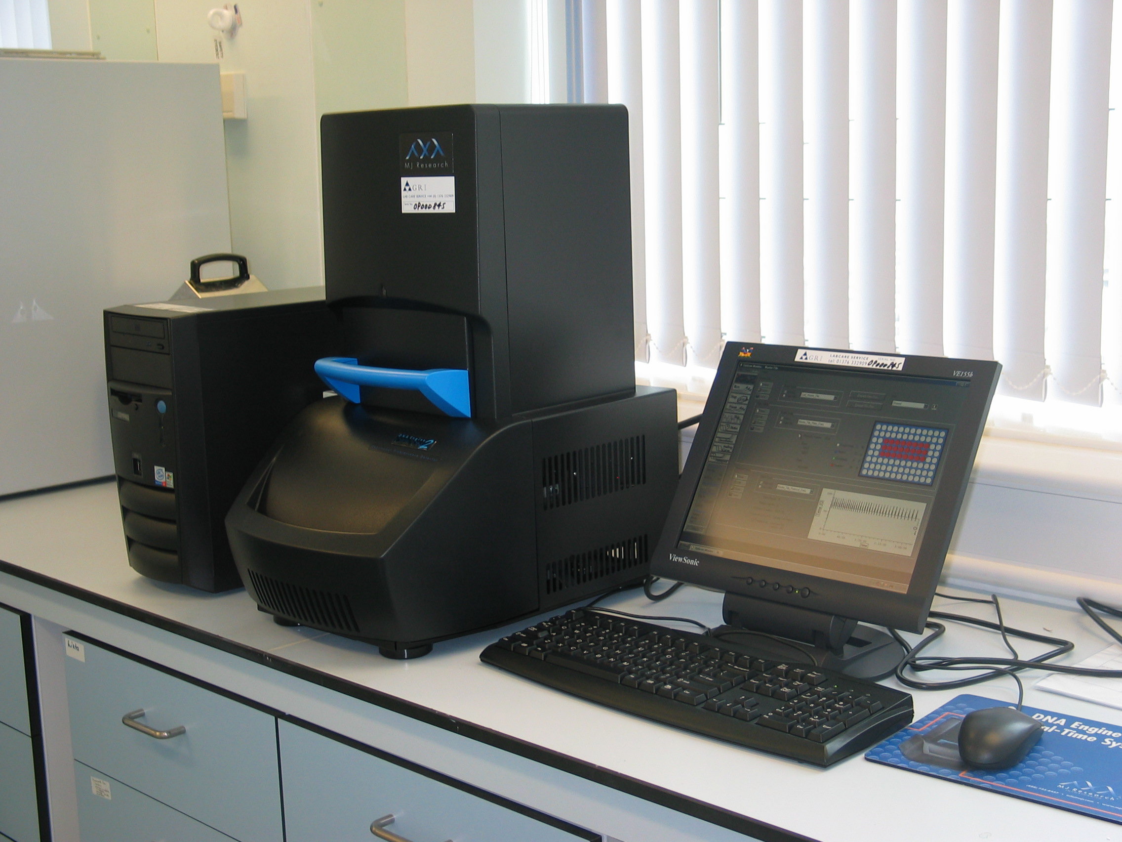
The Opticon system can be used to accurately quantify the initial concentration of a template in a sample.
In a quantitative experiment, samples of known concentration are run, then a standard curve is generated
by graphing the logarithm of known quantities vs. the threshold cycle for those samples.
Quantification of unknowns is performed by determining the amplification cycle at which the measured
fluorescence exceeds the background (i.e. the threshold cycle, or “C(t)”).
When the C(t) is applied to the standard curve generated from the known samples,
the initial concentration of the unknowns can be calculated to a high degree of accuracy.
For further details see this link
Read
a review about real-time PCR
Confocal Imaging of
living Neurones
We are using Leica SP
confocal systems based on the upright fixed-stage microscope with water
immersion lenses. We have established protocols which allow to visualise various
population of neurones targeted using viral vectors expressing different
fluorescent proteins in the same experiment. Organotypic slice cultures provide
an excellent model for this kind of approach.
BACK
TO HOMEPAGE