Dr Michael Knight, formally of the School of Psychological Science, used an Early Career Fellowship from the Elizabeth Blackwell Institute to model the physical mechanisms behind ageing disease processes and strokes using Magnetic Resonance Imaging (MRI), physical biochemistry and software engineering.
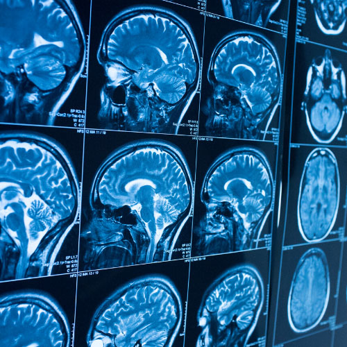
Dr Knight developed ‘relaxation anisotropy MRI imaging’ as a tool for investigating the brain - this essentially uses the processes by which the MRI signals change over time to increase the amount of information the MRI can extract. By understanding the criteria by which this relaxation anisotropy alters in brain disease states, it should be possible to increase the usefulness of MRI equipment in a variety of different ways, for example in understanding ageing, dementia assessment, and stroke onset diagnosis specific to stokes brought on by restriction in blood supply to tissue (known as ischaemia).
Dr Knight recruited 40 people from 23 to 71 years old, with no known brain disorders, and performed a range of MRI scans including the different modifications and analyses, and compared them to the computational models for anisotropy. Subtle but widespread changes as the brain ages could be detected, which raises the intriguing notion that different changes might be detectable in disease states, giving a much earlier diagnosis for e.g. Alzheimer’s disease than is currently possible.
Next, Dr Knight turned his attention to ischaemia brain injury and stroke. There is a specific therapeutic window in stroke treatment, beyond which it can be dangerous to administer many pharmaceutical treatments. However, in many cases the patients’ subjective reporting of symptom onset isn’t clear. In other words, if the patient can’t say when their symptoms began with any certainty, then many treatments will be denied them. Being able to retrospectively determine with certainty the time of stroke onset would therefore be of huge therapeutic advantage.
To date, Dr Knight has used anisotropy MIR imaging and computer modelling with a rat model of stroke to accurately determine the onset of ischaemia; the next step is to assess the feasibility of using the technique on patients with acute brain injury.
The Elizabeth Blackwell Institute funding proved extremely useful in progressing this work, said Dr Knight:“The project was very challenging by design, which has allowed me to develop as a scientist. In seeking to implement large-scale analyses, I was able to learn new computing and programming skills. I also became more familiar with various statistical tools as well as the magnetic resonance physics and mathematics required for the theoretical work."
"I had the opportunity to translate the work to different fields. This is another string to my bow that I would not have had without the fellowship; I have new collaborators and a new field!”